Image Credit: University of Adelaide
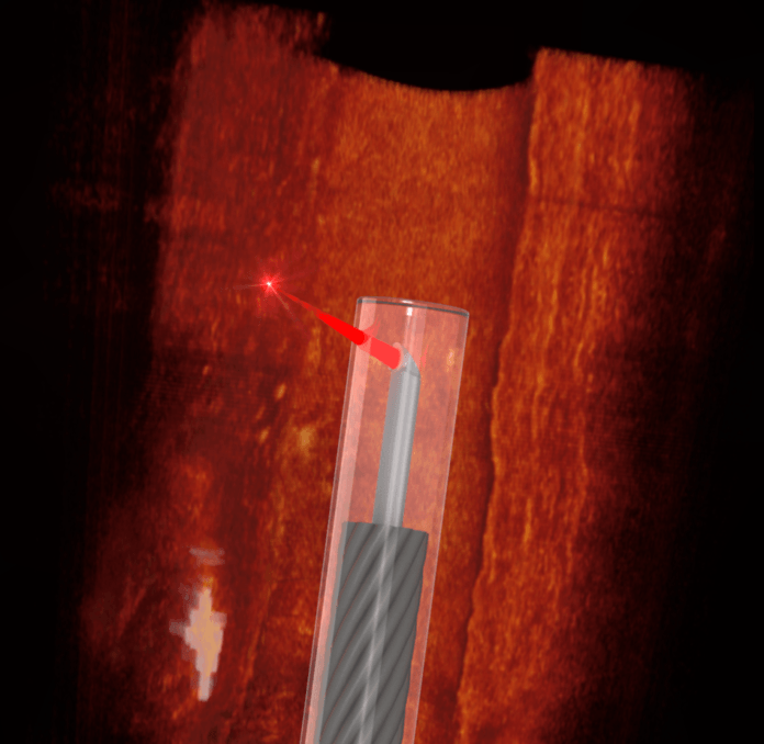
An optical probe with the thickness of a human hair, has been developed in a joint research effort by Australian and German medical clinicians and engineers. Named as the ‘smallest freeform 3D imaging probe’, it has proved its ability by 3D scanning from inside human and mice blood vessels.
In simpler terms, it is an ‘OCT scanning device’ for tiny spaces like blood vessels. But for the first time ever, scientists are able to get a clearer picture inside a blood vessel and be informed of plaques, fat/ cholesterol deposits or other substances which lead to numerous cardiovascular diseases.
The making of the ‘impossible’
Scientists first selected a 0.457mm wide optical fibre including the catheter sheath and micro-printed a tiny lens onto the fibre, facing sideways. Hard to see through the naked eye, this lens is less than 0.13mm in diameter. Then for the functioning part, they attached this tiny unit to an optical coherence tomography (OCT). OCT is known to map the retina in optometry. And the actual 3D scanning is done by a near-infrared light which goes through the tissue and buildup live high-resolution 3D images. Once inside a cavity, it can be rotated and pulled backwards slowly in order to get the full 3D map which will be 0.5mm deep from the contact surface. It is five times deeper than previous similar attempts. And this method ensures minimal trauma to delicate tissues, which is often an issue when using conventional endoscope insertion.
According to the study published in the Nature, Light Science & Applications, this has the potential to take unprecedented 3D scanning of tiny spaces in the body where conventional reach is impossible like the nervous system. And this will undoubtedly open doors for novel methods of minimally invasive applications in disease.

