Credit: www_slon_pics
A team of scientists found a certain molecule in our cells behaving abnormally in growing tumours that cause a rare liver cancer called, “Fibrolamellar carcinoma (FLC)”. This molecule is called “cyclic AMP” usually acting as a second messenger on glycogen around the liver.
The researchers used an array of modern techniques including, CRISPR gene editing, biosensor technology with fluorescent tools to observe these tiny molecules inside cells.
According to Professor Susan Taylor at the UC San Diego, “With the discovery of this new non-canonical mechanism for sequestering cAMP we build on the long-standing tradition of Roger Tsien (the late UC San Diego chemist and Nobel Prize winner), who engineered some of the first fluorescent probes that allowed us to query events taking place in the cell. Clearly the story of cAMP signalling still has new chapters,”
About Fibrolamellar Carcinoma (FLC)
It is a rare form of liver cancer affecting teens and people under 40s. The difference is that FLC normally affects people with healthy livers that do not have any damage from alcohol, drugs or certain viral infections. It takes up to 5% of all liver cancers.
The cell environment
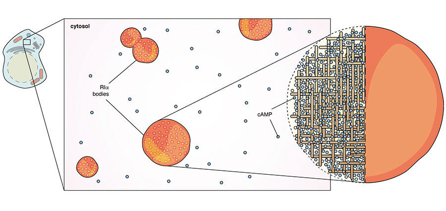
A living cell has organelles inside. Most of these organelles such as the mitochondria which generate energy are covered with a wall or membrane that protects the functions inside. But in this study scientists were amazed to notice a binding protein that connects to cAMP can form organelles without walls, i.e. they are membrane-less.
So, what happens with these membrane-less organelles?
According to Taylor, “We also found that cAMP is dynamically sequestered into these membrane-less organelles. And when these structures are destroyed, cAMP floods the cell, leading to uncontrolled cell growth—an event that can lead to tumour formation, our findings have provided a long-sought-after answer to the cAMP mystery.”

The main triggering point in the study was observing these membrane-less organelles (R1a bodies) gather together and dissociate apart. They noticed that these organelles “absorb” about 99% of all cAMPs in a cell. And these organelles occupy just about 1% of the cell. So this was a trigger for further examinations.
And they noticed that these cAMP filled organelles were interrupted by the major tumour causing fusion protein in Fibrolamellar carcinoma (FLC). And interestingly, this unexpected observation was, the reason behind the FLC’s tumour-causing mechanism, Eureka!
cAMPs are present inside membrane-less organelles not only in the liver but also in the tissue cells of the brain, heart and pancreas. This is something the researchers plan to study on in the future.
“As various membrane-less organelles formed in biological systems have attracted a lot of recent attention, a future direction is to look at what else is inside these structures and understanding the rules that allow molecules to enter and stay in these membrane-less organelles, related to cancer, we are actively looking into the molecular basis by which uncontrolled cAMP leads to cancer,” says, Professor Susan Taylor.
The study has been published in the journal Cell.

